How Do We Evaluate Varicose Veins?
Proper evaluation is essential for diagnosing and managing varicose veins, especially in advanced cases like venous ulcers. A structured approach involving history, clinical examination, imaging, and classification ensures optimal treatment.
History Taking
A detailed history provides critical insights into the progression of venous disease. Key aspects include:
- Symptoms: Patients may report heaviness, swelling, pain, itching, or night cramps. Symptoms often worsen after prolonged standing and improve with elevation.
- Ulcer History: Chronicity, recurrence, previous treatments, and healing response. Venous ulcers often have a long-standing history, unlike arterial ulcers.
- Risk Factors:
- Family history of varicose veins.
- Previous deep vein thrombosis (DVT) or trauma.
- Pregnancy, obesity, or prolonged standing occupation.
- Past venous interventions.
- Associated Skin Changes: Hyperpigmentation, lipodermatosclerosis, eczema, or atrophie blanche suggest chronic venous insufficiency.
Physical Examination
A. Limb Examination (Performed in Supine and Standing Positions)
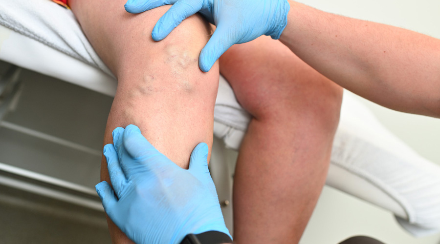
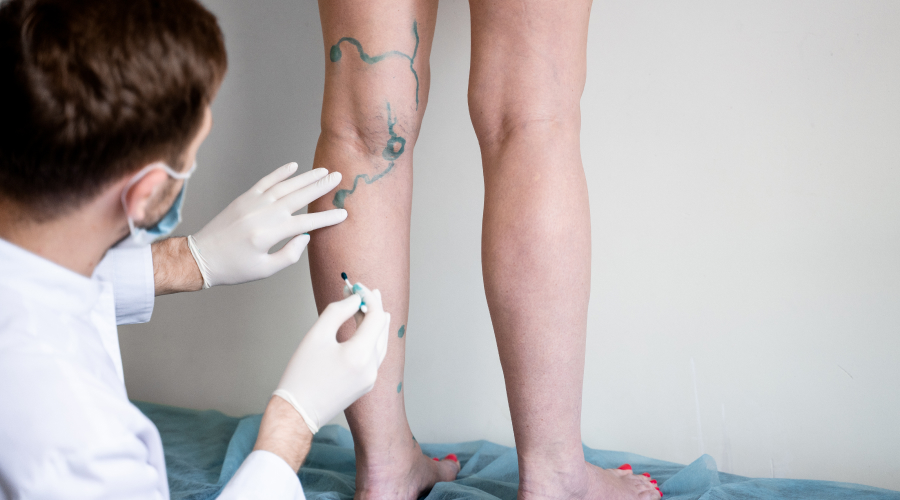
1. Inspection
- Assess the entire limb to visualize the extent of varicose veins.
- Identify the type of edema: pitting (venous insufficiency) vs. non-pitting (lymphedema).
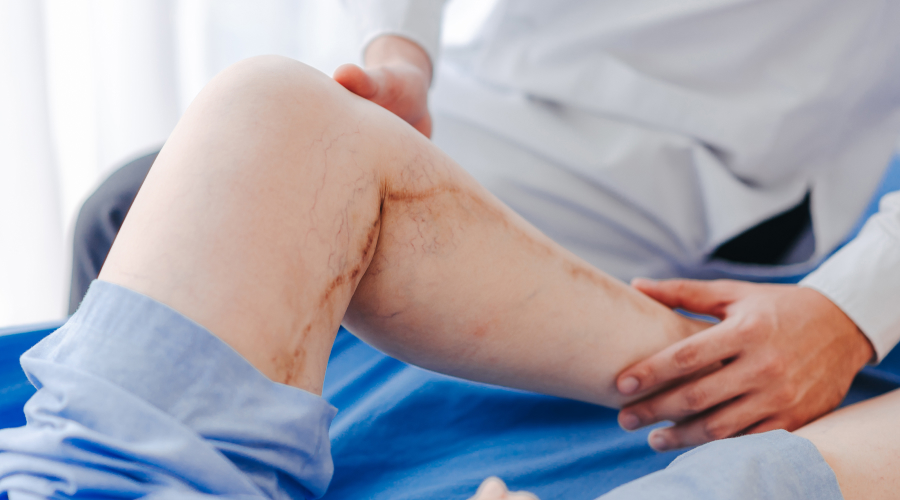
- Look for venous eczema, hyperpigmentation, lipodermatosclerosis, and atrophie blanche.
- Evaluate the presence of hemosiderin deposits or induration around the ankle.
2. Palpation:
- Assess the entire limb to visualize the extent of varicose veins.
- Identify the type of edema: pitting (venous insufficiency) vs. non-pitting (lymphedema).
- Look for venous eczema, hyperpigmentation, lipodermatosclerosis, and atrophie blanche.
- Evaluate the presence of hemosiderin deposits or induration around the ankle.
B. Venous Ulcer Evaluation
1. Location
- Venous ulcers are most commonly found over the medial malleolus (gaiter region).
- Less commonly, lateral malleolus or anterior shin may be involved.
2. Ulcer Characteristics
- Margins: Irregular and sloping edges.
- Base: Shallow with granulation tissue, may have fibrinous slough.
- Base: Shallow with granulation tissue, may have fibrinous slough.
3. Surrounding Skin
- Hyperpigmentation due to hemosiderin deposition.
- Lipodermatosclerosis (woody induration).
- Stasis dermatitis (venous eczema with erythema and scaling).
- Atrophie blanche (white scar-like patches).
4. Pain Assessment
- Typically mild to moderate pain, worsened by dependency and relieved by elevation.
- Severe pain suggests superadded infection or arterial involvement.
5. Signs of Infection
- Increased warmth, erythema, foul-smelling discharge.
- Presence of cellulitis or secondary bacterial colonization.
Ultrasound Doppler Examination
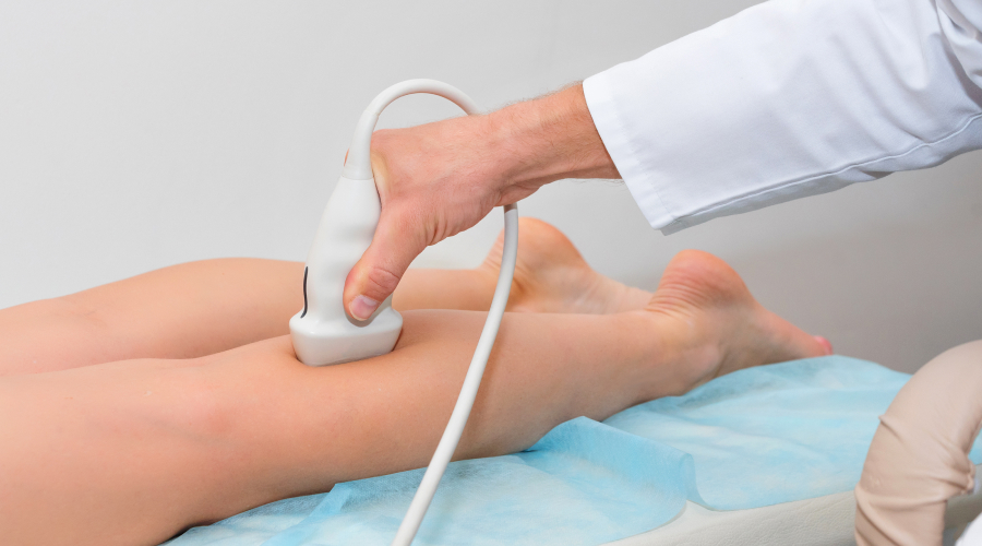
Color Doppler ultrasound (CDUS) is the gold standard for evaluating venous reflux and ulcer-related venous insufficiency.
- Superficial Venous Reflux: Great saphenous vein (GSV), small saphenous vein (SSV), and perforators.
- Deep Venous Reflux or Obstruction: To rule out post-thrombotic syndrome (PTS).
- Perforator Vein Incompetence: Perforators >3.5 mm with reflux are significant.
Advanced Imaging in Chronic Ulcers
If deep venous obstruction or iliac vein compression is suspected:
- CT Venography (CTV) or MR Venography (MRV): To evaluate May-Thurner Syndrome or venous outflow obstruction and pelvic congestion syndrome in non-healing ulcers.
Planning the Treatment
Treatment focuses on ulcer healing, preventing recurrence, and addressing underlying venous reflux.