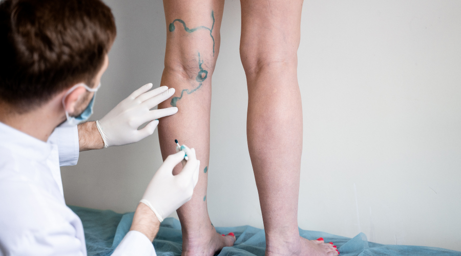
What Are Varicose Veins?
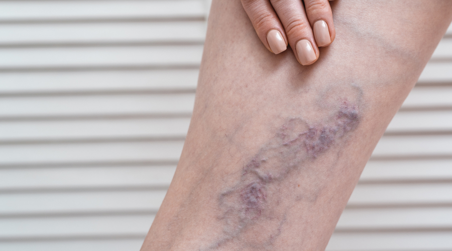
Varicose veins (VVs) are dilated, tortuous subcutaneous veins caused by venous valve insufficiency and blood reflux. They are part of chronic venous disease (CVD), which affects 2%-73% of adults globally. In India, the prevalence is approximately 30% of adults, with higher rates among healthcare workers and individuals in occupations requiring prolonged standing1,2,6. VVs can progress to complications such as venous ulcers and deep vein thrombosis (DVT), significantly impacting quality of life 3,5.
Etiology
The primary mechanisms contributing to varicose veins include:
- Valvular Insufficiency: Dysfunctional valves fail to prevent blood reflux, leading to pooling of blood in the veins1,6.
- Venous Wall Remodeling: Increased blood pressure stretches vein walls, weakening them and damaging valves6
Genetic Predisposition: Genome-wide association studies (GWAS) have identified genetic loci linked to vascular development7.
Risk Factors
Key modifiable and non-modifiable risk factors include

Age: Prevalence increases with age, affecting 50% of individuals over 50 years.

Sex: Females have a 2–3x higher risk due to hormonal influences and pregnancy.

Obesity: BMI over 30 increases risk by 30%.

Pregnancy: Multiparity elevates risk by 20%-40%.

Height: Taller stature is associated with increased risk (OR: 1.74 per 10 cm increase).

Prolonged Standing/Immobility: Occupations requiring standing for more than six hours per day increase risk by 60%.
In India, approximately 36.7% of cases are linked to prolonged standing occupations, particularly among healthcare workers.
Age Group
- <20 years: Rare (<5%) unless congenital2.
- 20–40 years: Prevalence rises to 10%-20%, often linked to pregnancy4.
- >50 years: Affects 30%-50%, with progressive venous wall degeneration6.
Sex Predilection
- Female Predominance: Approximately 60%-70% of cases are female, driven by hormonal changes and pregnancy-related venous pressure24.
- Male Prevalence: Accounts for about 20%-30%, often associated with occupational factors6.

Symptoms
Common symptoms include:
⦿Visible, veins that are bluish-purple or red in color.
⦿Pain, swelling, heaviness, aching, or burning sensations in the legs.
⦿Persistent edema (30%), skin pigmentation (20%), lipodermatosclerosis (15%).
⦿Asymptomatic cases account for approximately 20%-30% of patients.
Associated Medical Conditions
Varicose veins are associated with several medical conditions:
⦿ Deep Vein Thrombosis (DVT): Patients with VVs have a fivefold increased risk of DVT1,6.
⦿Peripheral Arterial Disease: An odds ratio of 1.5 is reported for patients with VVs3.
⦿Diabetes/Neuropathy: Accelerates venous ulcer progression (Hazard Ratio: 2.1)5.
Stages of Varicose Veins (CEAP Classification)
The CEAP classification system divides chronic venous disorders into seven clinical classes:
| Stage | Clinical Features | Subcategories/Details |
|---|---|---|
| C0 | No visible or palpable signs | - |
| C1 | Telangiectasias or reticular veins | - |
| C2 | Varicose veins | - |
| C3 | Edema due to venous etiology | - |
| C4a | Pigmentation and/or eczema | Red/brown discoloration; itchy skin; early inflammatory changes2,5,6 |
| C4b | Lipodermatosclerosis and/or atrophie blanche | Hardened/thickened skin; scar-like white patches; advanced inflammatory changes5,7 |
| C5 | Healed venous ulcer | - |
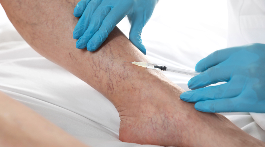
| C6 | Active venous ulcer | Open ulcers; requires surgical intervention |
Stage: C0
Clinical Features: No visible or palpable signs
Details: -
Stage: C1
Clinical Features: Telangiectasias or reticular veins
Details: -
Stage: C2
Clinical Features: Varicose veins
Details: -
Stage: C3
Clinical Features: Edema due to venous etiology
Details: -
Stage: C4a
Clinical Features: Pigmentation and/or eczema
Details: Red/brown discoloration; itchy skin; early inflammatory changes2,5,6
Stage: C4b
Clinical Features: Lipodermatosclerosis and/or atrophie blanche
Details: Hardened/thickened skin; scar-like white patches; advanced inflammatory changes5,7
Stage: C5
Clinical Features: Healed venous ulcer
Details: -
Stage: C6
Clinical Features: Active venous ulcer
Details: Open ulcers; requires surgical intervention
Detailed Subclassification for C4a and C4b
C4a
- Skin changes include pigmentation or eczema.
- Pigmentation often appears as brown or grey patches due to hemosiderin deposition.
- Eczema manifests as red, itchy skin caused by chronic inflammation.
C4b
Lipodermatosclerosis involves thickened or hardened skin due to chronic inflammation. Atrophie blanche refers to scar-like white patches surrounded by hyperpigmented areas.
- Both subcategories are markers of advanced venous disease progression146.
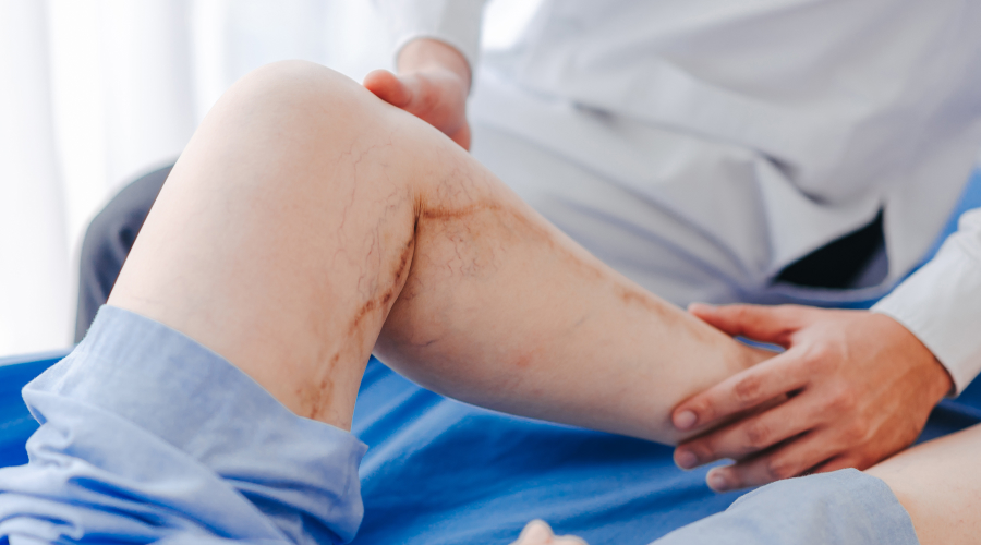
Corona Phlebectatica
Corona phlebectatica is classified under stage C4c in recent updates:
Features:
- Blue/red telangiectatic clusters around ankles ("crown-like" pattern).
- Stasis spots and perivenous pigmentation.
Grading:
- Grade I (Incipient): >5 non-confluent intradermal veins; limited distribution (<50% foot length).
- Grade II (Definite): Tortuous veins affecting ≥50% foot length.
Clinical Significance:
- Corona phlebectatica indicates advanced superficial + perforator reflux in up to 87% of cases57.
Corona Phlebectatica
Corona phlebectatica is classified under stage C4c in recent updates:
Features:
- Blue/red telangiectatic clusters around ankles ("crown-like" pattern)5,7..
Clinical Significance:
- Corona phlebectatica indicates advanced superficial + perforator reflux in up to 87% of cases.
References
1.Advancements in Varicose Vein Treatment (PMC). Published January 2024.
2.Varicose Veins Diagnosis and Treatment (American Family Physician). Published June 2019.
3.Varicose veins: Causes, symptoms, treatment (Medical News Today). Published December 2024.
4.The Basics of Varicose Veins (WebMD). Published April 2024.
5.The 100 most-cited articles on chronic venous disease (PMC). Published April 2020.
6.Varicose Veins Overview (Hopkins Medicine).
7.A Comprehensive Review on Varicose Veins (PubMed).
This updated overview using the provided references from PubMed-indexed journals and trusted medical sources.